|
Hướng dẫn tra cứu “Focus” hiệu quả bệnh do ký sinh trùng-cơ quan đích trong cơ thể người
Để đáp ứng yêu cầu bạn đọc về cách tra cứu “Focus” nhanh và hiệu quả bệnh so ký sinh trùng trong cơ thể người, Ban Biên tập Website xin cung cấp một Bảng hướng dẫn để bạn đọc quan tâm cùng tham khảo. Nhân một câu hỏi của bạn đọc (nguyenthehoa79@gmail....) ở thành phố Hồ Chí Minh đề cập và trao đổi về cách làm thế nào để nghĩ đến các tổn thương cơ quan đích do loại ký sinh trùng nào một cách tập trung và nhanh nhất để khỏi mất thời gian khi chúng tôi phát hiện các thương tổn, nhất là các hình ảnh viêm mô tế bào, hình ảnh abces có tróng âm, kèm theo hỗn hợp âm trên gan, lách, thận, mắt,… Chúng tôi rất cảm ơn bạn đã đưa ra một câu hỏi hay không những cho chúng tôi mà cho tất cả các anh chị và bạn bè đồng nghiệp cùng chia sẻ thông tin này để định hướng hay tập trung hơn để xác định ca bệnh một cách tối ưu nhất, nhanh nhất và tuân theo tiêu chí là chẩn đoán sớm và điều trị kịp thời. Nhân đây chúng tôi xin đưa ra một bảng hướng dẫn nhằm focus nhanh nhất và hình thành các bước tra cứu nhanh nhất để giúp bệnh nhân sớm khỏi bệnh và có một số ví dụ đi kèm. A. Một số loại ký sinh trùng thường gặp trên cơ quan tiêu hóa ở người 1. Esophagus ·Spirocerca lupi: oEggs [1] [2] [3] oDiagram [Adult] oLesions [Esophagus] [Aorta] [Aorta and esophagus] oAssociated lesions [Osteosarcoma] [Pulmonary hypertrophic osteoarthropathy] ·Gongylonema spp: oAdults [Showing cuticular plaques] oLesion [Ruminant esophagus] 2. Stomach, Rumen or Abomasum ·Habronema sp: (Draschia similar) oTypical lesion[Horse stomach] ·Physaloptera spp: oEggs [1] [2] [3] [4] oAdults [Head] [Male] [Male tail] [Female] oMorphology [Diagram of head] oLesion [Stomach of dog] ·Paramphistomum: oEgg [1] oAdult [Stained fluke] oLesion [Cyst in trachea] ·Ollulanus tricuspis oAdults [Adult male] [Adult female] oDiagram [Adult features] ·Ascarops (right) and Physocephalus (left) [Diagram of cephalic and oral structures] ·Gnathostoma [Diagram of head] 3. PGE Complex (trichostrongyle species found in the abomasum and small intestines of domestic ruminants) Trichostrongyloidea ·Typical eggs [1][2] [3] ·Trichostrongyle size comparison [Trichostrongylus, Ostertagia, Haemonchus] ·Ostertagia ostertagi [Male bursa on left] [Cervicle papilliae] [Abomasal lesions] [Lesions] ·Haemonchus contortus [Male bursa on right] [Bursa] [Spicules] [Cervicle papilliae] [Bottlejaw] ·Hyostrongylus rubidis (in swine stomach; comparable to Haemonchus) ·Marshallagia marshalli (abomasum of ruminants comparable to Haemonchus) ·Trichostrongylus axei [Male spicules] [Size; top pair of worms] ·Trichostrongylus (other species in small intestines) ·Cooperia [Cuticular striations] ·Nematodirus spp omale [Male head and tail] [Bursa with fused spicule] [Fused tip of spicules] ofemale[Egg on right] [Egg] [Cephalic inflation] [Female tail spine] 4. Small Intestines Ancylostomatoidea (Hookworms) ·Ancylostoma [Bursate tail] [Bursate tail] [Buccal capsule] [Scanning EM] [Ventral teeth diagram] ·Ancylostoma [Buccal capsule with ventral teeth] [Adult male and female Ancylostoma] ·Ancylostoma [Lesions in small intestines] [Cutaneous larval migrans (CLM)] [Histopath] ·Uncinaria spp and Bunostomum spp [Buccal capsule with ventral cutting plates] [Drawing of head] ·Hookworm eggs [Typical egg] [Egg] ·Globocephalus (pig hookworm with ventral cutting plates similar to Uncinaria) ·Necator americanus [Head of human hookworm] Ascaridoidea (Ascarids) ·Ascarid morphologic features [Adult male and female] [Lip diagram] [Ascarid lips] ·Toxocara canis eggs (Baylisascaris and T. vitulorum are similar) [Typical egg] [Larvated egg] ·Toxocara canis [Identifying features] [Adult ascarids in intestines] [Lung lesions] ·Toxascaris leonina [Typical Egg] [Egg] [Eggs:Toxocara, top; Toxascaris, bottom] [Adults in cat bile duct] ·Parascaris equorum [Typical egg] [Egg] [Adult ascarids in horse intestines] ·Ascaris suum [Egg] [Egg] [Liver with milk spots][Liver with milk spots] ·Ascaridia (Ascarid of poultry and other birds) Rhabditoidea ·Strongyloides (papillosus, ransomi, westeri) [Typical larvated egg] [Egg] [Egg] [Adults: small intestine histopath] ·Strongyloides (canis, stercoralis, tumefaciens) [Infective L3] [Infective L3] ·Strongyloides morphology [Diagram of stages] [Strongyloides L3 and Ancylostoma L3 compared] Trichinelloidea ·Capillaria bovis [Typical egg] [Stichosome with stichocytes] ·Trichinella spiralis [Drawing of adult male and female] [Female] [Female] [Male] [Tail of male] [Larva] ·Eggs size [Ancylostoma, Trichuris and Capillaria compared] Cestodes (Order Cyclophyllidea) Taeniidae: ·Eggs [Taeniid Egg] [Eggs] [Eggs] ·Metacestodes [Cysticercus][Cysticercus][Cysticerci in muscle][Cysticerci in viscera] ·Taenia spp [Adult morphology] [Scolex] [Circle of hooks] [Scolices] [Strobila] [Mature proglottids] ·Taeniiasis [Adult taeniids in small intestines of a dog] [Adults in gut] ·Echinococcus granulosus [Adult worm] [Hydatid cyst] [Hydatid cyst section] [Protoscolices] Anoplocephalidae: ·Eggs [Egg] [Egg] ·Metacestodes [Cysticercoid] ·Adults [Typical scolex] [Moniezia adult from cattle or sheep] [Anoplocephala in horse gut] Mesocestoididae [Egg] [Scolex] [Mature proglottids of Mesocestoides with paruterine organ] [Adult drawing] Dilepididae ·Dipylidium caninum [Egg packets] [Egg packets] ·Dipylidium caninum [Scolex] [Mature proglottids] [Gravid dried proglottid from bed sheets] Hymenolepididae [Hymenolepis diminuta egg] [Scolex] [Hymenolepis nana in mouse gut] Cestodes (Order Pseudophyllidea) Trematodes Acanthocephalans 5. Large intestines Strongyloidea Trichuroidea Oxyuroidea (pinworms) 6. Body Cavities and Viscera Body cavity Kidneys or Urinary bladder Liver - Dicrocoelium dentriticum (Family Dicrocoelidae - aquired from infected ants)[Adult fluke]
- Platynosomum fastosum(Family Dicrocoelidae - aquired from infected lizards) [Adult fluke]
- Eurytrema prycyononis (Family Dicrocoelidae - aquired from infected fish)
- Eurytrema pancreaticum (Family Dicrocoelidae - aquired from infected arthropods)
- Fasciola hepatica (Family Fasciolidae - metacercariae on vegetation)
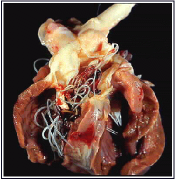
7. Cardiovascular and Respiratory Systems Cardiovascular system Parasites infecting the heart Parasite | Type of organism | Classification | Usual Hosts | Taenia saginata | Cestode | Taeniidae | Cattle (IH) | Taenia solium | Cestode | Taeniidae | Pigs (IH) | Neospora caninum | Protozoan | Apicomplexa | Dogs, others | Hepatozoon americanum | Protozoan | Apicomplexa | Dogs, others | Sarcocystis | Protozoan | Apicomplexa | Ruminants, others | Toxoplasma gondii | Protozoan | Apicomplexa | Dogs, cats (DH), others | Trypanosoma cruzi | Protozoan | Kinetoplastida | Dogs, other |
Dirofilaria immitis [Diagnostic microfilarial stage] [Microfilaria of Dipetalonema reconditum] [Adults in heart] [Intermediate host for heartworms] 9. Respiratory System 9. Helminth Parasites of the Somatic and Connective Tissues Skin and Connective Tissues Muscles Central Nervious System Eyes - Cysticercosis (Cysticercus cellulosae)
- Loa loa (human eye worm)
- Thelazia spp.
- Toxocara canis larvae
- Toxoplasma gondii
Trong phần này, nhiều tác giả phân loại đã rất chú ý đến các khía cạnh của từng loại ký sinh trùng mà chúng ta có thể nhìn thấy và có thể tra cứu một cách từ đầu đến cuối và có liên quan đến các bệnh lý khác gần với nó. Ngoài ra, các giai đoạn của bệnh của từng loại ký sinh trùng cũng như chu kỳ sinh bệnh cũng được trình bày nếu chúng ta gạch dưới chúng và tự tra cứu qua mạng internet sẽ có ngay một bài đầy đủ như trong sách giáo khoa. Bệnh có thể truyền từ động vật sang người và ngược lại. Bài viết gồm có tất cả các loại ký sinh trùng có thể chúng ta đã gặp và loại chưa bao giờ nghe đên hoặc chúng chỉ tồn tại ở châu Phi và một số quốc gia khác.
|


![[1]](http://instruction.cvhs.okstate.edu/jcfox/htdocs/Disk1/images/Img0044.jpg)
![[2]](http://instruction.cvhs.okstate.edu/jcfox/htdocs/Disk1/images/Img0044a.jpg)
![[3]](http://instruction.cvhs.okstate.edu/jcfox/htdocs/DISK1/IMAGES/spirocercaeggs.JPG)
![[Adult]](http://instruction.cvhs.okstate.edu/jcfox/htdocs/DISK1/IMAGES/Spirocerdgm.JPG)
![[Esophagus]](http://instruction.cvhs.okstate.edu/jcfox/htdocs/Disk1/images/lupinodule.jpg)
![[Aorta]](http://instruction.cvhs.okstate.edu/jcfox/htdocs/Disk1/images/lupiaorta.jpg)
![[Aorta and esophagus]](http://instruction.cvhs.okstate.edu/jcfox/htdocs/DISK1/IMAGES/osteosarcoma.JPG)
![[Osteosarcoma]](http://instruction.cvhs.okstate.edu/jcfox/htdocs/DISK1/IMAGES/Img0044c.jpg)
![[Showing cuticular plaques]](http://instruction.cvhs.okstate.edu/jcfox/htdocs/Disk1/images/Img0044d.jpg)
![[Ruminant esophagus]](http://instruction.cvhs.okstate.edu/jcfox/htdocs/Disk1/images/gongy2.jpg)
![[Horse stomach]](http://instruction.cvhs.okstate.edu/jcfox/htdocs/Disk1/images/Img0044e.jpg)
![[1]](http://instruction.cvhs.okstate.edu/jcfox/htdocs/Disk1/images/Img0043.jpg)
![[2]](http://instruction.cvhs.okstate.edu/jcfox/htdocs/Disk1/images/Img0043a.jpg)
![[3]](http://instruction.cvhs.okstate.edu/jcfox/htdocs/Disk1/images/Img0043b.jpg)
![[4]](http://instruction.cvhs.okstate.edu/jcfox/htdocs/Disk1/images/Img0043c.jpg)
![[Head]](http://instruction.cvhs.okstate.edu/jcfox/htdocs/DISK1/IMAGES/Img0068a1.JPG)
![[Male]](http://instruction.cvhs.okstate.edu/jcfox/htdocs/DISK1/IMAGES/physalopter1.JPG)
![[Male tail]](http://instruction.cvhs.okstate.edu/jcfox/htdocs/DISK1/IMAGES/Img0067a.JPG)
![[Female]](http://instruction.cvhs.okstate.edu/jcfox/htdocs/DISK1/IMAGES/physalopter2.JPG)
![[Diagram of head]](http://instruction.cvhs.okstate.edu/jcfox/htdocs/DISK1/IMAGES/Physalopdgm.JPG)
![[Stomach of dog]](http://instruction.cvhs.okstate.edu/jcfox/htdocs/Disk1/images/Img0043d.jpg)
![[1]](http://instruction.cvhs.okstate.edu/jcfox/htdocs/Disk1/images/Img0028b.jpg)
![[Stained fluke]](http://instruction.cvhs.okstate.edu/jcfox/htdocs/DISK1/IMAGES/Paramphistomum-1.jpg)
![[Cyst in trachea]](http://instruction.cvhs.okstate.edu/jcfox/htdocs/Disk1/Images/Img0088b.jpg)
![[Adult male]](http://instruction.cvhs.okstate.edu/jcfox/htdocs/Disk1/images/Ollulanus.jpg)
![[Adult features]](http://instruction.cvhs.okstate.edu/jcfox/htdocs/DISK1/IMAGES/oullanus.JPG)
![[Diagram of cephalic and oral structures]](http://instruction.cvhs.okstate.edu/jcfox/htdocs/DISK1/IMAGES/Ascarop_Physo.JPG)
![[Diagram of head]](http://instruction.cvhs.okstate.edu/jcfox/htdocs/DISK1/IMAGES/Gnathostdgm.JPG)
![[1]](http://instruction.cvhs.okstate.edu/jcfox/htdocs/Disk1/images/Img0045b.jpg)
![[2]](http://instruction.cvhs.okstate.edu/jcfox/htdocs/Disk1/images/Img0045f.jpg)
![[3]](http://instruction.cvhs.okstate.edu/jcfox/htdocs/Disk1/images/pseudo6.jpg)
![[Trichostrongylus, Ostertagia, Haemonchus]](http://instruction.cvhs.okstate.edu/jcfox/htdocs/Disk1/images/trichsize.jpg)
![[Male bursa on left]](http://instruction.cvhs.okstate.edu/jcfox/htdocs/Disk1/images/Img0064.jpg)
![[Cervicle papilliae]](http://instruction.cvhs.okstate.edu/jcfox/htdocs/Disk1/images/Img0072a1.jpg)
![[Abomasal lesions]](http://instruction.cvhs.okstate.edu/jcfox/htdocs/Disk1/images/Img0072d.jpg)
![[Lesions]](http://instruction.cvhs.okstate.edu/jcfox/htdocs/Disk1/images/Img0072e.jpg)
![[Bursa]](http://instruction.cvhs.okstate.edu/jcfox/htdocs/Disk1/images/Img0064a.jpg)
![[Spicules]](http://instruction.cvhs.okstate.edu/jcfox/htdocs/Disk1/images/Img0064a1.jpg)
![[Cervicle papilliae]](http://instruction.cvhs.okstate.edu/jcfox/htdocs/Disk1/images/Img0072a2.jpg)
![[Bottlejaw]](http://instruction.cvhs.okstate.edu/jcfox/htdocs/DISK1/IMAGES/bottlejaw.JPG)
![[Male spicules]](http://instruction.cvhs.okstate.edu/jcfox/htdocs/Disk1/images/Img0064aa.jpg)
![[Size; top pair of worms]](http://instruction.cvhs.okstate.edu/jcfox/htdocs/DISK1/IMAGES/trichsize.JPG)
![[Cuticular striations]](http://instruction.cvhs.okstate.edu/jcfox/htdocs/Disk1/images/Img0073.jpg)
![[Male head and tail]](http://instruction.cvhs.okstate.edu/jcfox/htdocs/Disk1/images/Img0073a1.jpg)
![[Bursa with fused spicule]](http://instruction.cvhs.okstate.edu/jcfox/htdocs/Disk1/images/Img0073a5.jpg)
![[Fused tip of spicules]](http://instruction.cvhs.okstate.edu/jcfox/htdocs/Disk1/images/Img0073a6.jpg)
![[Egg on right]](http://instruction.cvhs.okstate.edu/jcfox/htdocs/Disk1/images/Img0045.jpg)
![[Egg]](http://instruction.cvhs.okstate.edu/jcfox/htdocs/Disk1/images/Img0045c.jpg)
![[Cephalic inflation]](http://instruction.cvhs.okstate.edu/jcfox/htdocs/Disk1/images/Img0073a8.jpg)
![[Female tail spine]](http://instruction.cvhs.okstate.edu/jcfox/htdocs/Disk1/images/Img0073a9.jpg)
![[Bursate tail]](http://instruction.cvhs.okstate.edu/jcfox/htdocs/Disk1/images/Img0064aa3.jpg)
![[Bursate tail]](http://instruction.cvhs.okstate.edu/jcfox/htdocs/DISK1/IMAGES/Ancy_bursa3.JPG)
![[Buccal capsule]](http://instruction.cvhs.okstate.edu/jcfox/htdocs/Disk1/images/Img0069.jpg)
![[Scanning EM]](http://instruction.cvhs.okstate.edu/jcfox/htdocs/DISK1/IMAGES/ancyloemscan.jpg)
![[Ventral teeth diagram]](http://instruction.cvhs.okstate.edu/jcfox/htdocs/Disk1/images/Img0069a.jpg)
![[Buccal capsule with ventral teeth]](http://instruction.cvhs.okstate.edu/jcfox/htdocs/Disk1/images/Img0069a1.jpg)
![[Adult male and female Ancylostoma]](http://instruction.cvhs.okstate.edu/jcfox/htdocs/Disk1/images/hooks.jpg)
![[Lesions in small intestines]](http://instruction.cvhs.okstate.edu/jcfox/htdocs/Disk1/images/hookgut.jpg)
![[Cutaneous larval migrans (CLM)]](http://instruction.cvhs.okstate.edu/jcfox/htdocs/Disk1/images/Img0069b.jpg)
![[Histopath]](http://instruction.cvhs.okstate.edu/jcfox/htdocs/DISK1/IMAGES/ancylostomabite.JPG)
![[Buccal capsule with ventral cutting plates]](http://instruction.cvhs.okstate.edu/jcfox/htdocs/Disk1/images/Img0070.jpg)
![[Drawing of head]](http://instruction.cvhs.okstate.edu/jcfox/htdocs/Disk1/images/Img0070b.jpg)
![[Typical egg]](http://instruction.cvhs.okstate.edu/jcfox/htdocs/Disk1/images/Img0045a.jpg)
![[Head of human hookworm]](http://instruction.cvhs.okstate.edu/jcfox/htdocs/Disk1/images/Img0070a.jpg)
![[Adult male and female]](http://instruction.cvhs.okstate.edu/jcfox/htdocs/Disk1/images/Img0038a.jpg)
![[Lip diagram]](http://instruction.cvhs.okstate.edu/jcfox/htdocs/DISK1/IMAGES/asclips.JPG)
![[Ascarid lips]](http://instruction.cvhs.okstate.edu/jcfox/htdocs/Disk1/images/Img0077.jpg)
![[Typical egg]](http://instruction.cvhs.okstate.edu/jcfox/htdocs/Disk1/images/Img0035.jpg)
![[Larvated egg]](http://instruction.cvhs.okstate.edu/jcfox/htdocs/Disk1/images/Img0035c.jpg)
![[Identifying features]](http://instruction.cvhs.okstate.edu/jcfox/htdocs/DISK1/IMAGES/ascaridspp.jpg)
![[Adult ascarids in intestines]](http://instruction.cvhs.okstate.edu/jcfox/htdocs/Disk1/images/Img0038b.jpg)
![[Lung lesions]](http://instruction.cvhs.okstate.edu/jcfox/htdocs/DISK1/IMAGES/asclunglesion.jpg)
![[Typical Egg]](http://instruction.cvhs.okstate.edu/jcfox/htdocs/Disk1/images/Img0036.jpg)
![[Egg]](http://instruction.cvhs.okstate.edu/jcfox/htdocs/DISK1/IMAGES/Toxascarisegg.jpg)
![[Eggs:Toxocara, top; Toxascaris, bottom]](http://instruction.cvhs.okstate.edu/jcfox/htdocs/Disk1/images/Img0037.jpg)
![[Adults in cat bile duct]](http://instruction.cvhs.okstate.edu/jcfox/htdocs/Disk1/images/Img0037a.jpg)
![[Typical egg]](http://instruction.cvhs.okstate.edu/jcfox/htdocs/Disk1/images/Img0039.jpg)
![[Egg]](http://instruction.cvhs.okstate.edu/jcfox/htdocs/Disk1/images/Img0039b.jpg)
![[Adult ascarids in horse intestines]](http://instruction.cvhs.okstate.edu/jcfox/htdocs/Disk1/images/Img0039d.jpg)
![[Egg]](http://instruction.cvhs.okstate.edu/jcfox/htdocs/clinpara/homebanrjpg.jpg)
![[Egg]](http://instruction.cvhs.okstate.edu/jcfox/htdocs/Disk1/images/Img0038.jpg)
![[Liver with milk spots]](http://instruction.cvhs.okstate.edu/jcfox/htdocs/Disk1/images/Img0038c.jpg)
![[Liver with milk spots]](http://instruction.cvhs.okstate.edu/jcfox/htdocs/Disk1/images/Img0038c1.jpg)
![[Typical larvated egg]](http://instruction.cvhs.okstate.edu/jcfox/htdocs/Disk1/images/Img0046.jpg)
![[Egg]](http://instruction.cvhs.okstate.edu/jcfox/htdocs/Disk1/images/Img0046a.jpg)
![[Egg]](http://instruction.cvhs.okstate.edu/jcfox/htdocs/Disk1/images/Img0046b.jpg)
![[Adults: small intestine histopath]](http://instruction.cvhs.okstate.edu/jcfox/htdocs/Disk1/images/Img0047a.jpg)
![[Infective L3]](http://instruction.cvhs.okstate.edu/jcfox/htdocs/Disk1/images/Img0047.jpg)
![[Infective L3]](http://instruction.cvhs.okstate.edu/jcfox/htdocs/DISK1/IMAGES/Strongyloidesl3.JPG)
![[Diagram of stages]](http://instruction.cvhs.okstate.edu/jcfox/htdocs/DISK1/IMAGES/strgloi3stages.JPG)
![[Strongyloides L3 and Ancylostoma L3 compared]](http://instruction.cvhs.okstate.edu/jcfox/htdocs/Disk1/images/Img0048.jpg)
![[Typical egg]](http://instruction.cvhs.okstate.edu/jcfox/htdocs/Disk1/images/Img0041b.jpg)
![[Stichosome with stichocytes]](http://instruction.cvhs.okstate.edu/jcfox/htdocs/Disk1/images/Carole7.jpg)
![[Drawing of adult male and female]](http://instruction.cvhs.okstate.edu/jcfox/htdocs/Disk1/images/Img0079b.jpg)
![[Female]](http://instruction.cvhs.okstate.edu/jcfox/htdocs/Disk1/images/Img0079b2.jpg)
![[Female]](http://instruction.cvhs.okstate.edu/jcfox/htdocs/Disk1/images/Img0079b3.jpg)
![[Male]](http://instruction.cvhs.okstate.edu/jcfox/htdocs/Disk1/images/Img0079bb.jpg)
![[Tail of male]](http://instruction.cvhs.okstate.edu/jcfox/htdocs/Disk1/images/Img0079bbb.jpg)
![[Larva]](http://instruction.cvhs.okstate.edu/jcfox/htdocs/Disk1/images/Img0079e.jpg)
![[Taeniid Egg]](http://instruction.cvhs.okstate.edu/jcfox/htdocs/Disk1/Images/Img0031.jpg)
![[Eggs]](http://instruction.cvhs.okstate.edu/jcfox/htdocs/Disk1/Images/Img0031b.jpg)
![[Eggs]](http://instruction.cvhs.okstate.edu/jcfox/htdocs/Disk1/Images/Img0031c.jpg)
![[Cysticercus]](http://instruction.cvhs.okstate.edu/jcfox/htdocs/Disk1/Images/Img0031d.jpg)
![[Cysticercus]](http://instruction.cvhs.okstate.edu/jcfox/htdocs/Disk1/Images/Img0059f.jpg)
![[Cysticerci in muscle]](http://instruction.cvhs.okstate.edu/jcfox/htdocs/Disk1/Images/Img0059g.jpg)
![[Cysticerci in viscera]](http://instruction.cvhs.okstate.edu/jcfox/htdocs/Disk1/Images/Img0059h.jpg)
![[Adult morphology]](http://instruction.cvhs.okstate.edu/jcfox/htdocs/Disk1/Images/Img0059.jpg)
![[Scolex]](http://instruction.cvhs.okstate.edu/jcfox/htdocs/Disk1/Images/Img0059b.jpg)
![[Circle of hooks]](http://instruction.cvhs.okstate.edu/jcfox/htdocs/Disk1/Images/Img0059a.jpg)
![[Scolices]](http://instruction.cvhs.okstate.edu/jcfox/htdocs/Disk1/Images/Img0059b1.jpg)
![[Strobila]](http://instruction.cvhs.okstate.edu/jcfox/htdocs/Disk1/Images/Img0059c.jpg)
![[Mature proglottids]](http://instruction.cvhs.okstate.edu/jcfox/htdocs/Disk1/Images/Img0059d.jpg)
![[Adult taeniids in small intestines of a dog]](http://instruction.cvhs.okstate.edu/jcfox/htdocs/Disk1/images/taeniasis.jpg)
![[Adults in gut]](http://instruction.cvhs.okstate.edu/jcfox/htdocs/Disk1/Images/Img0059e.jpg)
![[Adult worm]](http://instruction.cvhs.okstate.edu/jcfox/htdocs/Disk1/Images/Img0059i.jpg)
![[Hydatid cyst]](http://instruction.cvhs.okstate.edu/jcfox/htdocs/Disk1/Images/Img0059j.jpg)
![[Hydatid cyst section]](http://instruction.cvhs.okstate.edu/jcfox/htdocs/Disk1/Images/Img0059k.jpg)
![[Protoscolices]](http://instruction.cvhs.okstate.edu/jcfox/htdocs/Disk1/Images/protoscolices.jpg)
![[Egg]](http://instruction.cvhs.okstate.edu/jcfox/htdocs/Disk1/Images/Img0033.jpg)
![[Egg]](http://instruction.cvhs.okstate.edu/jcfox/htdocs/Disk1/Images/Img0033b.jpg)
![[Cysticercoid]](http://instruction.cvhs.okstate.edu/jcfox/htdocs/Disk1/Images/Img0062a.jpg)
![[Typical scolex]](http://instruction.cvhs.okstate.edu/jcfox/htdocs/Disk1/Images/Img0063a.jpg)
![[Moniezia adult from cattle or sheep]](http://instruction.cvhs.okstate.edu/jcfox/htdocs/Disk1/Images/Img0063.jpg)
![[Anoplocephala in horse gut]](http://instruction.cvhs.okstate.edu/jcfox/htdocs/Disk1/Images/Img0063b.jpg)
![[Egg]](http://instruction.cvhs.okstate.edu/jcfox/htdocs/Disk1/Images/Img0034.jpg)
![[Scolex]](http://instruction.cvhs.okstate.edu/jcfox/htdocs/Disk1/Images/Img0034a.jpg)
![[Adult drawing]](http://instruction.cvhs.okstate.edu/jcfox/htdocs/Disk1/Images/Img0034aa.jpg)
![[Egg packets]](http://instruction.cvhs.okstate.edu/jcfox/htdocs/Disk1/Images/Img0032.jpg)
![[Egg packets]](http://instruction.cvhs.okstate.edu/jcfox/htdocs/Disk1/Images/Img0032a.jpg)
![[Scolex]](http://instruction.cvhs.okstate.edu/jcfox/htdocs/Disk1/Images/Img0061.jpg)
![[Mature proglottids]](http://instruction.cvhs.okstate.edu/jcfox/htdocs/Disk1/Images/Img0060.jpg)
![[Gravid dried proglottid from bed sheets]](http://instruction.cvhs.okstate.edu/jcfox/htdocs/Disk1/Images/Img0062.jpg)
![[Hymenolepis diminuta egg]](http://instruction.cvhs.okstate.edu/jcfox/htdocs/Disk1/Images/Img0034b.jpg)
![[Scolex]](http://instruction.cvhs.okstate.edu/jcfox/htdocs/Disk1/Images/Img0063d.jpg)
![[Hymenolepis nana in mouse gut]](http://instruction.cvhs.okstate.edu/jcfox/htdocs/Disk1/Images/Img0063e.jpg)
![[Egg]](http://instruction.cvhs.okstate.edu/jcfox/htdocs/Disk1/Images/Img0030a.jpg)
![[Scolex]](http://instruction.cvhs.okstate.edu/jcfox/htdocs/Disk1/Images/Img0030b.jpg)
![[Mature proglottids]](http://instruction.cvhs.okstate.edu/jcfox/htdocs/Disk1/Images/Img0030c.jpg)
![[Egg]](http://instruction.cvhs.okstate.edu/jcfox/htdocs/Disk1/Images/Img0030.jpg)
![[1st Intermediate host]](http://instruction.cvhs.okstate.edu/jcfox/htdocs/Disk1/Images/Img0030d.jpg)
![[Typical egg with operculum]](http://instruction.cvhs.okstate.edu/jcfox/htdocs/Disk1/Images/Img0028.jpg)
![[Typical egg]](http://instruction.cvhs.okstate.edu/jcfox/htdocs/Disk1/Images/Img0028a.jpg)
![[Nanophyetes salmincola adult]](http://instruction.cvhs.okstate.edu/jcfox/htdocs/Disk1/Images/Img0089.jpg)
![[Adult fluke]](http://instruction.cvhs.okstate.edu/jcfox/htdocs/Disk1/Images/Img0089a.jpg)
![[Metacercariae in fish]](http://instruction.cvhs.okstate.edu/jcfox/htdocs/Disk1/Images/Img0089b.jpg)
![[Opisthorchis species similar to Amphimeris pseudofelineus in cats]](http://instruction.cvhs.okstate.edu/jcfox/htdocs/Disk1/Images/Img0092.jpg)
![[Echinostoma adult]](http://instruction.cvhs.okstate.edu/jcfox/htdocs/Disk1/Images/Img0092a.jpg)
![[Alaria adult]](http://instruction.cvhs.okstate.edu/jcfox/htdocs/Disk1/Images/Img0093.jpg)
![[Strigea adult]](http://instruction.cvhs.okstate.edu/jcfox/htdocs/Disk1/Images/Img0093a.jpg)
![[Egg]](http://instruction.cvhs.okstate.edu/jcfox/htdocs/Disk1/Images/Img0029.jpg)
![[Egg]](http://instruction.cvhs.okstate.edu/jcfox/htdocs/Disk1/Images/Img0029a.jpg)
![[Egg]](http://instruction.cvhs.okstate.edu/jcfox/htdocs/Disk1/Images/Img0029a1.jpg)
![[Egg]](http://instruction.cvhs.okstate.edu/jcfox/htdocs/Disk1/Images/Img0029a2.jpg)
![[Egg]](http://instruction.cvhs.okstate.edu/jcfox/htdocs/Disk1/Images/Img0029aa.jpg)
![[Thorny proboscis]](http://instruction.cvhs.okstate.edu/jcfox/htdocs/Disk1/Images/Img0029c.jpg)
![[Thorny proboscis]](http://instruction.cvhs.okstate.edu/jcfox/htdocs/Disk1/Images/Img0029d.jpg)
![[Adult worms attached to small intestine]](http://instruction.cvhs.okstate.edu/jcfox/htdocs/Disk1/Images/Img0029b.jpg)
![[Adult worm found in can of tuna fish]](http://instruction.cvhs.okstate.edu/jcfox/htdocs/Disk1/Images/Img0029e.jpg)
![[Egg]](http://instruction.cvhs.okstate.edu/jcfox/htdocs/Disk1/Images/Img0045.jpg)
![[Eggs]](http://instruction.cvhs.okstate.edu/jcfox/htdocs/Disk1/Images/Img0045d.jpg)
![[Eggs]](http://instruction.cvhs.okstate.edu/jcfox/htdocs/Disk1/Images/Img0045e.jpg)
![[Eggs]](http://instruction.cvhs.okstate.edu/jcfox/htdocs/Disk1/Images/Img0045e1.jpg)
![[Bursate tail]](http://instruction.cvhs.okstate.edu/jcfox/htdocs/Disk1/Images/Img0064aa3.jpg)
![[Buccal capsule]](http://instruction.cvhs.okstate.edu/jcfox/htdocs/Disk1/images/Img0071a.jpg)
![[Anterior mesenteric artery]](http://instruction.cvhs.okstate.edu/jcfox/htdocs/Disk1/images/Img0071b.jpg)
![[Intestinal infarct]](http://instruction.cvhs.okstate.edu/jcfox/htdocs/Disk1/images/colicgut.jpg)
![[Adult male]](http://instruction.cvhs.okstate.edu/jcfox/htdocs/DISK1/IMAGES/Strongylus1.JPG)
![[Assorted adults]](http://instruction.cvhs.okstate.edu/jcfox/htdocs/Disk1/images/Img0071.jpg)
![[Buccal capsule]](http://instruction.cvhs.okstate.edu/jcfox/htdocs/Disk1/images/Img0071c.jpg)
![[Buccal capsule]](http://instruction.cvhs.okstate.edu/jcfox/htdocs/Disk1/images/Img0071cc.jpg)
![[Buccal capsule]](http://instruction.cvhs.okstate.edu/jcfox/htdocs/Disk1/images/Img0071d.jpg)
![[Buccal capsule]](http://instruction.cvhs.okstate.edu/jcfox/htdocs/Disk1/images/Img0071e.jpg)
![[Buccal capsule]](http://instruction.cvhs.okstate.edu/jcfox/htdocs/Disk1/images/Img0071e2.jpg)
![[Buccal capsule]](http://instruction.cvhs.okstate.edu/jcfox/htdocs/Disk1/images/Img0071e3.jpg)
![[Nodular lesion]](http://instruction.cvhs.okstate.edu/jcfox/htdocs/Disk1/images/Img0071f.jpg)
![[Adult]](http://instruction.cvhs.okstate.edu/jcfox/htdocs/Disk1/images/Img0065.jpg)
![[Adult diagram]](http://instruction.cvhs.okstate.edu/jcfox/htdocs/DISK1/IMAGES/Trichurisdgm.jpg)
![[Egg]](http://instruction.cvhs.okstate.edu/jcfox/htdocs/Disk1/Images/Img0040.jpg)
![[Egg]](http://instruction.cvhs.okstate.edu/jcfox/htdocs/Disk1/Images/Img0040a.jpg)
![[Egg]](http://instruction.cvhs.okstate.edu/jcfox/htdocs/Disk1/Images/Img0040b.jpg)
![[Adults]](http://instruction.cvhs.okstate.edu/jcfox/htdocs/Disk1/Images/Img0074.jpg)
![[Adults in large intestines]](http://instruction.cvhs.okstate.edu/jcfox/htdocs/Disk1/Images/Img0074a.jpg)
![[Rectal prolapse in human child]](http://instruction.cvhs.okstate.edu/jcfox/htdocs/DISK1/IMAGES/prolapse.JPG)
![[Egg]](http://instruction.cvhs.okstate.edu/jcfox/htdocs/Disk1/Images/Img0042.jpg)
![[Pinworm diagram]](http://instruction.cvhs.okstate.edu/jcfox/htdocs/DISK1/IMAGES/pinworm1.jpg)
![[Adult female]](http://instruction.cvhs.okstate.edu/jcfox/htdocs/Disk1/Images/Img0078.jpg)
![[Adult females]](http://instruction.cvhs.okstate.edu/jcfox/htdocs/Disk1/Images/Oxyurisfe.jpg)
![[Adult female]](http://instruction.cvhs.okstate.edu/jcfox/htdocs/DISK1/IMAGES/pinworm.JPG)
![[Adult with immature Strongylus edentatus found in peritoneal cavity of a zebra]](http://instruction.cvhs.okstate.edu/jcfox/htdocs/Disk1/images/setaria_strongylus.jpg)
![[Egg]](http://instruction.cvhs.okstate.edu/jcfox/htdocs/Disk1/images/Img0054.jpg)
![[Egg]](http://instruction.cvhs.okstate.edu/jcfox/htdocs/Disk1/images/Img0054a.jpg)
![[Egg]](http://instruction.cvhs.okstate.edu/jcfox/htdocs/Disk1/images/Img0054c.jpg)
![[Adult]](http://instruction.cvhs.okstate.edu/jcfox/htdocs/Disk1/Images/Img0054d.jpg)
![[Drawing of male bursa]](http://instruction.cvhs.okstate.edu/jcfox/htdocs/Disk1/Images/Img0054e.jpg)
![[Adult male and females]](http://instruction.cvhs.okstate.edu/jcfox/htdocs/Disk1/images/stephanurus.jpg)
![[Kidney lesion]](http://instruction.cvhs.okstate.edu/jcfox/htdocs/Disk1/images/kidney.jpg)
![[Adults]](http://instruction.cvhs.okstate.edu/jcfox/htdocs/Disk1/Images/Img0094.jpg)
![[Adult fluke]](http://instruction.cvhs.okstate.edu/jcfox/htdocs/Disk1/Images/Img0091.jpg)
![[Adult fluke]](http://instruction.cvhs.okstate.edu/jcfox/htdocs/Disk1/Images/Platynosomum.jpg)
![[Fasciola hepatica egg]](http://instruction.cvhs.okstate.edu/jcfox/htdocs/Disk1/Images/Img0028b.jpg)
![[Fasciola hepatica adult]](http://instruction.cvhs.okstate.edu/jcfox/htdocs/Disk1/Images/Img0087a.jpg)
![[Lymnied snail in typical habitat]](http://instruction.cvhs.okstate.edu/jcfox/htdocs/Disk1/Images/Img0087c1.jpg)
![[Fascioloides magna adult]](http://instruction.cvhs.okstate.edu/jcfox/htdocs/Disk1/Images/Img0087b.jpg)
![[Liver lesions]](http://instruction.cvhs.okstate.edu/jcfox/htdocs/Disk1/Images/capillarialiver.jpg)
![[Histopath of liver lesion filled with eggs]](http://instruction.cvhs.okstate.edu/jcfox/htdocs/Disk1/Images/Img0041a.jpg)
![[Protoscolices in hydatid cyst]](http://instruction.cvhs.okstate.edu/jcfox/htdocs/Disk1/Images/protoscolicies.jpg)
![[Protoscolices similar to those in alveolar cyst]](http://instruction.cvhs.okstate.edu/jcfox/htdocs/Disk1/Images/prodoscolicies.jpg)
![[Adult scolex]](http://instruction.cvhs.okstate.edu/jcfox/htdocs/Disk1/Images/Img0063c1.jpg)
![[Fringed proglottids]](http://instruction.cvhs.okstate.edu/jcfox/htdocs/Disk1/Images/Img0063c2.jpg)
![[Fringed proglottids]](http://instruction.cvhs.okstate.edu/jcfox/htdocs/Disk1/Images/Img0063c3.jpg)
![[Diagnostic microfilarial stage]](http://instruction.cvhs.okstate.edu/jcfox/htdocs/Disk1/Images/Img0052.jpg)
![[Microfilaria of Dipetalonema reconditum]](http://instruction.cvhs.okstate.edu/jcfox/htdocs/Disk1/Images/Img0053.jpg)
![[Adults in heart]](http://instruction.cvhs.okstate.edu/jcfox/htdocs/Disk1/Images/Img0052a.jpg)
![[Intermediate host for heartworms]](http://instruction.cvhs.okstate.edu/jcfox/htdocs/Disk1/Images/mosquito.jpg)
![[Elephantiasis]](http://instruction.cvhs.okstate.edu/jcfox/htdocs/DISK1/IMAGES/elephantiasis.JPG)
![[Diagnostic larval stage]](http://instruction.cvhs.okstate.edu/jcfox/htdocs/Disk1/Images/Img0051.jpg)
![[Worms in trachea]](http://instruction.cvhs.okstate.edu/jcfox/htdocs/Disk1/Images/Img0051c.jpg)
![[Worms in trachea]](http://instruction.cvhs.okstate.edu/jcfox/htdocs/Disk1/Images/Img0051d.jpg)
![[Diagnostic larval stage]](http://instruction.cvhs.okstate.edu/jcfox/htdocs/Disk1/Images/Img0051a.jpg)
![[Diagnostic egg stage]](http://instruction.cvhs.okstate.edu/jcfox/htdocs/Disk1/Images/Img0050.jpg)
![[Lung lesions similar to Muellerius]](http://instruction.cvhs.okstate.edu/jcfox/htdocs/Disk1/Images/Img0051b.jpg)
![[Egg]](http://instruction.cvhs.okstate.edu/jcfox/htdocs/Disk1/images/Img0041e.jpg)
![[Bursa and spicules of male]](http://instruction.cvhs.okstate.edu/jcfox/htdocs/Disk1/Images/Img0049b.jpg)
![[Diagnostic larval stage]](http://instruction.cvhs.okstate.edu/jcfox/htdocs/Disk1/Images/Img0049.jpg)
![[L3 attached to fungal spore]](http://instruction.cvhs.okstate.edu/jcfox/htdocs/Disk1/Images/Img0049f.jpg)
![[Worms in trachea of cow]](http://instruction.cvhs.okstate.edu/jcfox/htdocs/Disk1/Images/Img0049e.jpg)
![[Infected lungs]](http://instruction.cvhs.okstate.edu/jcfox/htdocs/DISK1/IMAGES/dictyosheep.JPG)
![[Diagnostic larval stage]](http://instruction.cvhs.okstate.edu/jcfox/htdocs/Disk1/Images/Img0049a.jpg)
![[Worms in trachea]](http://instruction.cvhs.okstate.edu/jcfox/htdocs/Disk1/Images/Img0049d.jpg)
![[Adult]](http://instruction.cvhs.okstate.edu/jcfox/htdocs/Disk1/Images/Img0088.jpg)
![[Second intermediate host]](http://instruction.cvhs.okstate.edu/jcfox/htdocs/Disk1/Images/Img0088a.jpg)
![[Copepod intermediate host]](http://instruction.cvhs.okstate.edu/jcfox/htdocs/DISK1/IMAGES/copepod2.jpg)
![[Lesion]](http://instruction.cvhs.okstate.edu/jcfox/htdocs/Disk1/images/ochocerca_lesion.jpg)
![[Lesion on human]](http://instruction.cvhs.okstate.edu/jcfox/htdocs/Disk1/images/onchocerca_head.jpg)
![[Skin lesion caused by Stephanofilaria]](http://instruction.cvhs.okstate.edu/jcfox/htdocs/Disk1/images/Stephan2.jpg)
![[Male]](http://instruction.cvhs.okstate.edu/jcfox/htdocs/DISK1/IMAGES/trichinellamale.JPG)
![[Female]](http://instruction.cvhs.okstate.edu/jcfox/htdocs/DISK1/IMAGES/trichinellafe.JPG)
![[Drawing of Trichinella male and femal]](http://instruction.cvhs.okstate.edu/jcfox/htdocs/Disk1/Images/Img0079b.jpg)
![[Encysted larvae]](http://instruction.cvhs.okstate.edu/jcfox/htdocs/Disk1/Images/Img0079c.jpg)
![[Encysted larvae]](http://instruction.cvhs.okstate.edu/jcfox/htdocs/Disk1/Images/Img0079d.jpg)
![[Neural dirofilariasis]](http://instruction.cvhs.okstate.edu/jcfox/htdocs/Disk1/Images/Img0052a1.jpg)